Patient was. 30/M, known RHD, presented with
Acute onset SOB GR IV NYHA
chest pain
Palpitations.
On clinical examination,
Pulse 120bpm regular rhythm (sinus tachycardia)
BP 90/60 mm Hg
JVP FULL
B/L minimal pitting pedal edema
CVS
apex L 5th ICS inside MCL tapping type
PALPABLE THRILL AT MITRAL AREA
DIASTOLIC SHOCK (palpable P2) at PA
Palpable thrill at tricuspid area
LOUD P2 at Pulmonary area,
SOFT S1 at Mitral area
MDM GR IV at MA ,low pitched,rumbling murmur, best heard with bell in Left lateral position at expiration.
PSM GR IV at TA ,high pitched , best heard in supine position at end expiration
EDM at NAA Gr II high pitched, best heard I'm sitting position at end expiration.
ECG FINDINGS
Bi Atrial enlargement…
RV +
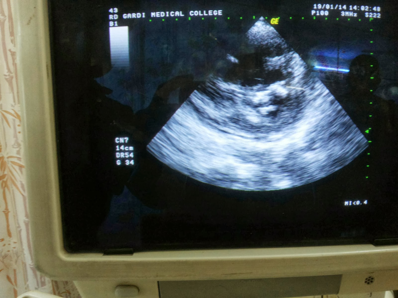
ECHOCARDIOGRAPHY FINDINGS
Heavily calcified Mitral Valve leaflets
Severe MS
MVA 0.66 cm ²( PHT method)
0.82cm ² ( PSAX Planimetry)
Grossly dilated RA &RV
RA volume approx 300 -350,ml
(77x70x71)
RVID basal Diameter 41mm
And now the most interesting finding
Severe TR WITH 'Double ' JET
I tried to see if the jets were arising fromthe same place, but on close observation and different planes & views, it was clear that both jets were different.
One jet arised from the normal TRICUSPID INFLOW while the other jet originated somewhere laterally .
The velocities of both these regurgitant jets were different.
Central jet being 4.5 m/s
While lateral jet 2.6m/s
They both gave a separate distinct jets visible on Color Doppler.
there was also mild PULMONARY REGURGITATION,
Mild MITRAL REGURGITATION
MILD AORTIC STENOSIS WITH MILD AORTIC INCOMPETENCE.
thankfully there were no clots.
FINAL DIAGNOSIS :
VHD
severe MS (?CRITICAL) with heavily calcified both leaflets
mild MR
Grossly dilated RA RV
SEV TR WITH DOUBLE JET (? TRICUSPID LEAFLET PERFORATION)
SEV PAH
(PASP 75 mm Hg)
Mild AS with AR.
Probably Rheumatic in origin


No comments:
Post a Comment